
Lameness exams
The team at Gibbs Marsh have extensive experience in lameness investigations, poor performance and sports horse medicine. Whether your horse is struggling at the end of a busy season, on three legs or just not performing to the standard you would expect, we will help you get to the bottom of your concerns.
Vicky O'Gorman holds dedicated clinic days at Gibbs Marsh to work up the lame or poor-performing horse. Here at the clinic we have all of the facilities and tools needed, as well as highly experienced vet technicians to work with your horse for the best diagnostic and therapeutic results.
We are also able to come out to your yard. If you would like to bring your horse to us, please call the office to see when the next clinic day is due to be held.

A thorough lameness examination is a structured process, beginning with a detailed conversation about your concerns and expectations for your horse's future.
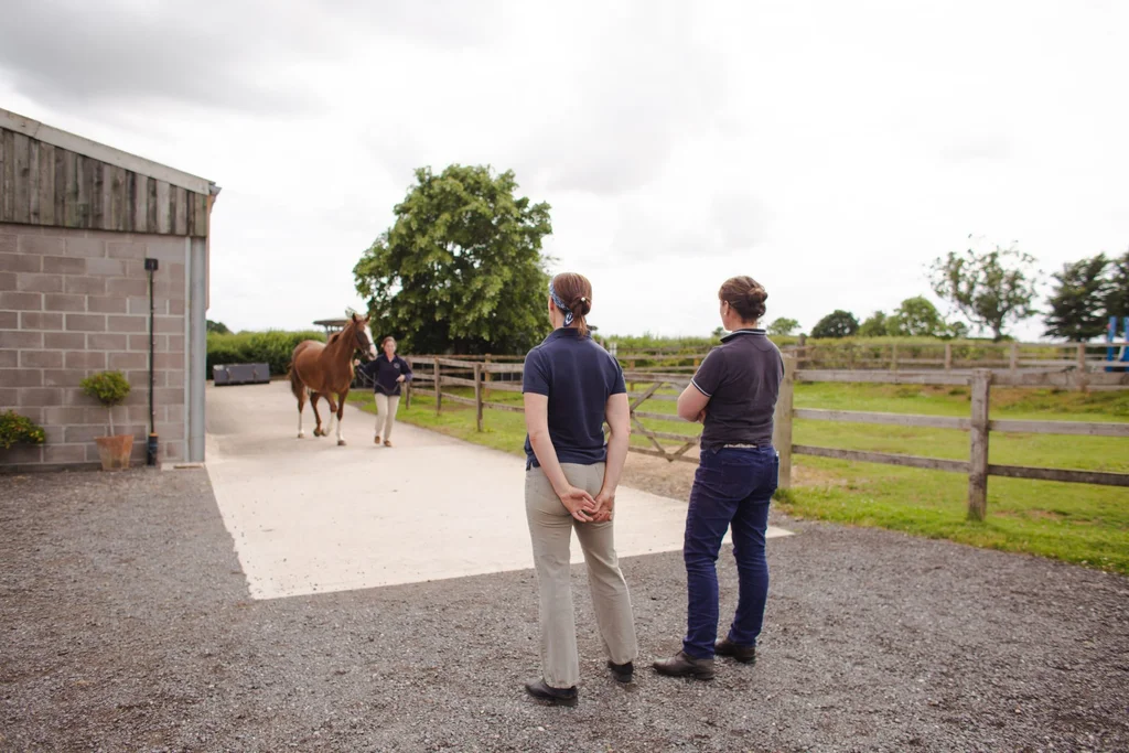
We will assess your horse in walk, trot and canter on the straight and on the circle, as appropriate. We have a hard trot-up, a non-slip hard lunging area and an arena to see the horse moving on multiple surfaces. We are also able to assess the horse under saddle in the school if necessary. Palpation of the whole horse can help to detect swelling, heat and pain in a limb, guiding us in narrowing down the source of the lameness.
Flexion tests are often performed following an initial trot-up and help us evaluate how the manipulation of certain joints affects the subsequent movement of the horse. Flexions are a subjective assessment, however they frequently help to clarify the presence of a low-grade lameness as well as indicating the location of pain in the limb.
Nerve blocks are one of our best aids in objectively identifying sources of pain. They involve the injection of small volumes of local anaesthetic into joints, or around specific nerves that innervate regions of a limb, to numb their sensation. We generally begin at the foot and work our way up the limb, repeating the trot-up after each block to assess any change in the lameness.
Once the vet has narrowed down the source of pain, a multitude of diagnostic aids may be appropriate for further investigation, including radiography and ultrasound scanning of the affected area.

